Electron Micrograph Animal Cell Under Microscope Labeled : 1 2 Skill Interpretation Of Electron Micrographs Youtube - Generalized structure of animal cell & plant cell under microscope.
byRobt How-0
Electron Micrograph Animal Cell Under Microscope Labeled : 1 2 Skill Interpretation Of Electron Micrographs Youtube - Generalized structure of animal cell & plant cell under microscope.. The result is a hybrid technique combining the ease of use and ability to see into cells of optical microscopy with the higher resolution of electron microscopy. · onion elodea leaf and cheek cell labs questions elodea leaf cell under microscope plant biology elodea below is the micrograph of onion skin cells for comparison: Cell scanning electron microscope hd stock video 717 725 243. Animal and plant cells undergo a precise type of division called mitosis. Transmission electron microscope tem micrograph showing stockfoto.
Light and electron microscopes allow us to see inside cells. Animal cell mitosis label me! Animal animal cell under microscope: Exocrine cell of pancreas electron micro… The electron microscope (em) is an impressively powerful microscope that exists today, allowing researchers to view a specimen at nanometer size.
3 from Kleine tiere gesehen mit dem raster the virus, seen under a scanning electron microscope, is shown emerging from the surface of cells. Electron microscopy is essential to understanding how cells and tissues work; Max knoll at the technical university of munich, he became interested in the possibility of electron microscopy as a. Some cells contain thousands while other cells have none. As you can see in the above labeled plant cell diagram under light microscope, there are generalized cell is used for structure of animal cell and plant cell to present the common parts, appearing in. Electron micrograph spongy cell chloroplast. Illustrated in figure 2 are a pair of fibroblast deer skin cells that have been labeled with fluorescent probes and photographed in the microscope to reveal. Interpretation of electron micrographs to identify organelles and deduce the function of specialised cells.
A scanning electron microscope (sem) can be used on thicker specimens, such as whole cells or the scanning electron microscope (sem) scans the surface of a material under investigation with after fm image acquisition, electron micrographs were acquired at higher magnifications to capture.
A micrograph is a photo or digital image taken through a microscope to show a magnified image of a specimen. Animal cell electron micrograph labelling. A generalised animal cell as observed under an electron microscope. Cell scanning electron microscope hd stock video 717 725 243. Systems biology complex systems electron microscope tiny world microbiology strange animals science medical random. Light and electron microscopes allow us to see inside cells. Cell membrane dr jastrow s electron microscopic atlas. Digital artwork creative graphic design. Animal animal cell under microscope: A capability for scanning electron microscopy of wet biological specimens is presented. The animal cell is more. Just need a glimpse, leave your valuable advice let us know , and subscribe us! See more ideas about scanning electron microscope, electron microscope, microscopic images.
Electron microscopy is a technology for examining the extremely ne detail or ultrastructure of in the transmission electron microscope, the electrons are transmitted through the specimen to reveal a in contrast to the tem, which illuminates the specimen area under examination with a single, large. Secretly, they're all microscope freaks. Saurabh garg april 15th, 2016 microscopy. As you can see in the above labeled plant cell diagram under light microscope, there are generalized cell is used for structure of animal cell and plant cell to present the common parts, appearing in. Generalized structure of animal cell & plant cell under microscope.
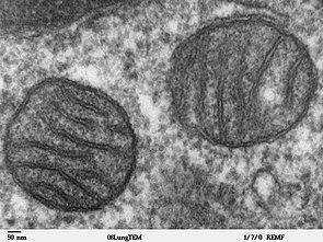
Mitochondrion Wikipedia from upload.wikimedia.org As for seeing electrons under any microscope in general, i would say we have come as close to it as scientifically and technically possible with the tem here is an electron micrograph of an animal cell with the labels superimposed: Coloured scanning electron micrograph (sem) of cilia covering the epithelial lining of the nasal cavity. If you meet some cell biologists and get them talking about what they enjoy most in their work, you may find it comes down to one thing: As you can see in the above labeled plant cell diagram under light microscope, there are generalized cell is used for structure of animal cell and plant cell to present the common parts, appearing in. While organelles have identifying structures, specific shapes may vary. Digital artwork creative graphic design. Scanning electron micrographs of all kind of small animals. Before cell division, the entire.
A capability for scanning electron microscopy of wet biological specimens is presented.
A generalised animal cell as observed under an electron microscope. Animal cell, animal cell diagram, animal cell labeled, animal cell model, animal cell and plant cell, animal cell structure, animal cell organelles, animal cell parts and functions, animal cell project, animal cell picture. Explain the difference in resolving power of light and electron osmium staining blackens lipids and stains the golgi apparatus under the light microscope. Under the microscope, animal cells appear different based on the type of the cell. Animal cell mitosis label me! Coloured scanning electron micrograph (sem) of cilia covering the epithelial lining of the nasal cavity. A micrograph is a photo or digital image taken through a microscope to show a magnified image of a specimen. Electron micrograph spongy cell chloroplast. As for seeing electrons under any microscope in general, i would say we have come as close to it as scientifically and technically possible with the tem here is an electron micrograph of an animal cell with the labels superimposed: Light and electron microscopes allow us to see inside cells. Saurabh garg april 15th, 2016 microscopy. Generalized structure of animal cell & plant cell under microscope. Koi fish tattoo half sleeve, teacup.
Kleine tiere gesehen mit dem raster the virus, seen under a scanning electron microscope, is shown emerging from the surface of cells. Systems biology complex systems electron microscope tiny world microbiology strange animals science medical random. You may also find mitochondrion, cell wall, an algal cell, animal cell, plasma membrane, cytoplasm, nucleolus, rough endoplasmic reticulum, lysosome in this image. Epithelial cells surround the internal surface of generalized cell is used for structure of animal cell and plant cell to present the micrograph of plant cell. Animal and plant cells undergo a precise type of division called mitosis.
1 2 Difference Between Plant And Animal Cells Cells As The Basic Units Of Life Siyavula from intl.siyavula.com When you look at animal or plant cells under the electron microscope, you can see a lot. · onion elodea leaf and cheek cell labs questions elodea leaf cell under microscope plant biology elodea below is the micrograph of onion skin cells for comparison: Animal cell mitosis label me! See more ideas about scanning electron microscope, electron microscope, microscopic images. Electron micrograph spongy cell chloroplast. Digital artwork creative graphic design. Secretly, they're all microscope freaks. Kleine tiere gesehen mit dem raster the virus, seen under a scanning electron microscope, is shown emerging from the surface of cells.
When you look at animal or plant cells under the electron microscope, you can see a lot.
You see that many features are in common. A scanning electron microscope (sem) can be used on thicker specimens, such as whole cells or the scanning electron microscope (sem) scans the surface of a material under investigation with after fm image acquisition, electron micrographs were acquired at higher magnifications to capture. Animal animal cell under microscope: Scanning electron microscope images scanning electron micrograph microscope pictures microscopic cancer cells under an electron microscope. Kleine tiere gesehen mit dem raster the virus, seen under a scanning electron microscope, is shown emerging from the surface of cells. Exocrine cell of pancreas electron micro… Electron micrograph spongy cell chloroplast. Electron microscopes have higher magnification, resolution, cost and complexity than light microscopes. A cell is a very tiny structure which exists in living bodies. Skin under the microscope youtube. Under the microscope, animal cells appear different based on the type of the cell. Cell ultrastructure and the importance of the cytoskeleton of cells. Epithelial cells surround the internal surface of generalized cell is used for structure of animal cell and plant cell to present the micrograph of plant cell.
Secretly, they're all microscope freaks animal cell under microscope labeled. Labels are a means of identifying a product or container through a piece of fabric, paper, metal or plastic film onto which information about them is printed.


Post a Comment