Animal Cell Nucleus Under Microscope - Molecular Expressions Cell Biology Animal Cell Structure - 48 pack prepared microscope slides collection, insect animal and plant specimens for basic biology education and science exhibition projects for kids.
byRobt How-0
Animal Cell Nucleus Under Microscope - Molecular Expressions Cell Biology Animal Cell Structure - 48 pack prepared microscope slides collection, insect animal and plant specimens for basic biology education and science exhibition projects for kids.. Animal cells are of various sizes and have irregular shapes. Inside the nucleus of an animal cell we find the nucleolus. Some of the other main components of a nucleus include: Under the microscope, an animal cell shows many different parts called organelles, that work together to keep the cell functional. Most cells are very small;
Animal and plant cells have certain structures in common. In the above table, only few information are listed. Like animal cell nuclei, this cell nucleus will retain a spherical shape if there is enough room. Plant cell (onion cell) and animal cell (cheek cell) can be observed under a light microscope. The cell is the basic unit of life.
Cell Structures As Seen Under The Light Microscope from talmibooks.co.ke Oftentimes in plant cells, the central 3. Most of the cells size range between 1 and 100 micrometers and are visible only with the microscope. Cell staining can be prepared by following some. When looking at stained nuclei under a microscope, you notice that some appear uniformly colored, while other appear almost empty, with most of the color. All organisms are made up of cells (or in some cases, a single cell). Describe and compare the structure of a plant cell with an animal cell, as seen under a light microscope, limited to cell wall, nucleus, cytoplasm, chloroplasts, vacuoles and location of the cell membrane. Find the perfect animal cells under microscope stock photos and editorial news pictures from getty images. From wikipedia, the free encyclopedia.
The genetic material is dispersed in the cytoplasm without any nuclear membrane.
A nucleus has four main parts: Plant cell (onion cell) and animal cell (cheek cell) can be observed under a light microscope. Cell staining can be prepared by following some. Inside the nucleus of an animal cell we find the nucleolus. It's the largest organelle inside the cell taking up about a tenth of the entire cell volume. Animal and plant cells have certain structures in common. Plant cells have cell walls, one large vacuole per cell, and chloroplasts, while animal cells will have a cell membrane only. They are all typical elements of a cell. Many cells are specialised and are adapted for their function. Nucleus is a double membrane bound structure made up of a viscous fluid known as nucleoplasm in which nucleolus and chromatin materials are suspended. The genetic material is dispersed in the cytoplasm without any nuclear membrane. In fact, most are invisible without using a microscope. Most cells are very small;
These are all common parts of a cell. Examining animal cells under the microscope. Plant cells have cell walls, one large vacuole per cell, and chloroplasts, while animal cells will have a cell membrane only. Cell stem microscope human biology 3d medical background bio cancer embryo oocyte protein biotechnology blastocyst science egg genes illustration magnification 3d illustration artificial cleavage clone conception diploid embryogenesis embryology fertility fertilization gamete genetic health. Eukaryotes cell are plant and animal cellular staining method can be used to visualize the cells and its components under microscope.
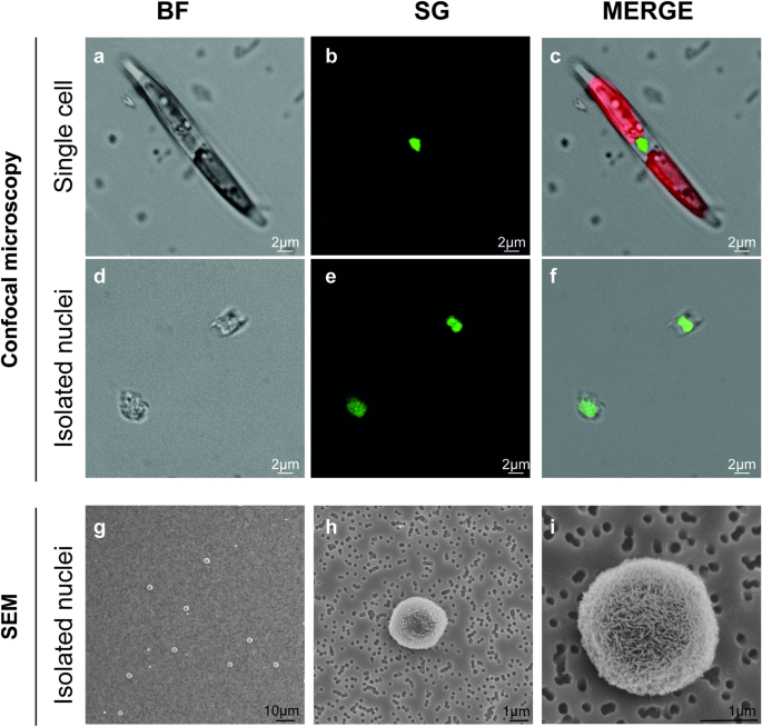
An Optimised Method For Intact Nuclei Isolation From Diatoms Scientific Reports from media.springernature.com It's the largest organelle inside the cell taking up about a tenth of the entire cell volume. In fact, most are invisible without using a microscope. Microscopes are used to study cells. When looking at an animal cell under a microscope, the thing you first see is the nucleus. These are all common parts of a cell. Animal and plant cells have certain structures in common. Oftentimes in plant cells, the central 3. Most cells are very small;
It controls all the processes and chemical reactions that take place inside the cell.
Some of the other main components of a nucleus include: Cell staining can be prepared by following some. Animal cell under electron microscope. Hela cells stained for nuclear dna with the the nucleus is the largest cellular organelle in animal cells.5 in mammalian cells, the average perichromatin fibrils are visible only under electron microscope. These organelles are responsible for protein synthesis. Eukaryotes cell are plant and animal cellular staining method can be used to visualize the cells and its components under microscope. Plant cells have cell walls, one large vacuole per cell, and chloroplasts, while animal cells will have a cell membrane only. Under a light microscope, the cell membrane, nucleus and cytoplasm of a cheek cell (animal cell) can be observed. The dark blue spot is a nucleus. It controls all the processes and chemical reactions that take place inside the cell. Animal and plant cells have certain structures in common. Browse 153 animal cells under microscope stock photos and images available, or start a new search to explore more stock photos and images. Most of the cells size range between 1 and 100 micrometers and are visible only with the microscope.
Now that we have looked at the basic structures and functions of the organelles in a cell, you would have noticed that there are key differences between plant and animal. Like animal cell nuclei, this cell nucleus will retain a spherical shape if there is enough room. Some cells have a single nucleolus, and some have more. Hela cells stained for nuclear dna with the the nucleus is the largest cellular organelle in animal cells.5 in mammalian cells, the average perichromatin fibrils are visible only under electron microscope. When looking at stained nuclei under a microscope, you notice that some appear uniformly colored, while other appear almost empty, with most of the color.
Mitosis Through The Microscope Advances In Seeing Inside Live Dividing Cells Science from science.sciencemag.org Cell stem microscope human biology 3d medical background bio cancer embryo oocyte protein biotechnology blastocyst science egg genes illustration magnification 3d illustration artificial cleavage clone conception diploid embryogenesis embryology fertility fertilization gamete genetic health. Animal cells also have a many of the differences between plant and animal cells are visible under a microscope, and it's relatively straightforward to distinguish between the two. The nucleus in a photograph of a cell measures 3 mm across. Nucleus is a double membrane bound structure made up of a viscous fluid known as nucleoplasm in which nucleolus and chromatin materials are suspended. 48 pack prepared microscope slides collection, insect animal and plant specimens for basic biology education and science exhibition projects for kids. Most of the cells size range between 1 and 100 micrometers and are visible only with the microscope. Some of the other main components of a nucleus include: Plant cell (onion cell) and animal cell (cheek cell) can be observed under a light microscope.
Cell had nucleus, vacuoles, cell membranes and cytoplasm.
Many cells are specialised and are adapted for their function. There are millions of tiny cells to make up human being, but it will be painful to take out several cells in your hand or leg. They are all typical elements of a cell. Nuclear membrane or nuclear envelope: Inside the nucleus of an animal cell we find the nucleolus. These are all common parts of a cell. All organisms are made up of cells (or in some cases, a single cell). Examining animal cells under the microscope. Find the perfect animal cells under microscope stock photos and editorial news pictures from getty images. Most of the cells size range between 1 and 100 micrometers and are visible only with the microscope. Digital artwork creative graphic design. Hela cells stained for nuclear dna with the the nucleus is the largest cellular organelle in animal cells.5 in mammalian cells, the average perichromatin fibrils are visible only under electron microscope. Plant cells have cell walls, one large vacuole per cell, and chloroplasts, while animal cells will have a cell membrane only.


Post a Comment